

Apex 4B
Apex SEC columns
Easily isolate pure, functional EVs with high yield


Apex SEC plates
Scale your isolation
- Get the same results as Apex SEC columns in a plate format.
- Compatible with the Summit instrument


Apex SEC Working Principle
Size-exclusion chromatography (SEC) gently separates extracellular vesicles (EVs) by size as a sample passes through a column packed with porous beads. Large particles that cannot enter the pores elute first, while smaller molecules (e.g., secreted proteins) enter the pores and travel a longer path, causing them to elute later. This creates a key trade-off: smaller pore sizes increase EV yield but allow more contaminants to co-elute, whereas larger pores improve purity by retaining more mid-sized impurities but reduce EV recovery.
Apex MM for lipoprotein depletion
More than just SEC
By combining SEC with a multi-mode (MM) resin that uses hydrophobic interaction and ion-exchange to selectively bind and remove lipoproteins and other soluble contaminants, Apex MM columns enhance EV purity.

| Sample Type | Analysis Type | Recommended Column |
|---|---|---|
| Plasma, Serum, or Cell Culture Media (with FBS/Lipoproteins) | Single Particle (e.g. NTA/Nanoflow) | Apex MM |
| Targeted Assays (e.g. ELISA/WB/ONI/Leprechaun) | Apex 4B | |
| Untargeted Assays (e.g. Proteomics) | Apex MM | |
| Cell Culture Media (Serum-free Media) | Most applications | Apex 6B |
| CSF | ||
| Urine |
How do I apply Apex SEC columns to my research?
Columns selector Table
Quickly identify the optimal SEC column for your workflow. This tool guides selection based on sample type and downstream analysis, helping you choose from Apex MM, Apex 4B, and Apex 6B columns. Built with expert input, the table distills technical guidance into an easy-to-use reference.

EV elution profile of 8 Apex 6B columns, which are optimized for consistent drip speed. CV (EV yield) =4.3%
| Column format | Column | Plate | mini |
|---|---|---|---|
| Column type | MM / 4B / 6B | 4B / 6B | MM |
| Input sample volume | 0.5 - 1.0 mL | 0.5 - 1.0 mL | 0.1 - 0.3 mL |
| Column volume | 9.0 mL | 9.0 mL | 3.0 mL |
| Column reproducibility | 10% CV | ||
| Sample types | plasma, serum, urine, CSF, cell culture media | ||
| Resin types | 4% or 6 % cross linked agarose beads, cross linked agarose plus multi-mode | ||
| Exclusion limit | 35 nm or 20 nm | ||
Technical information
The Apex size exclusion chromatography (SEC) column purifies extracellular vesicles (EVs) from biological fluids such as plasma, serum, urine, cell culture media, or cerebrospinal fluid (CSF).
There are Apex columns for your different sample types and volumes.
Discuss your isolation needs with our scientific team
Latest Apex Application Notes
Can SEC be a scalable and more reproducible alternative to ultracentrifugation?
Can SEC be a scalable and more reproducible alternative to ultracentrifugation?
Read More
Advancing Plasma-Derived Extracellular Vesicle Analysis Through High-Purity Isolation and Particle-Specific Quantification
Advancing Plasma-Derived Extracellular Vesicle Analysis Through High-Purity Isolation and Particle-Specific Quantification
Read More
Drip speed matters: Enhance EV isolation reproducibility with Apex columns and validate using the Atlas ELISA
Drip speed matters: Enhance EV isolation reproducibility with Apex columns and validate using the Atlas ELISA
Read More
Optimizing extracellular vesicle isolation: Why Apex MM columns outperform conventional SEC columns for isolating EVs from plasma or serum
Optimizing extracellular vesicle isolation: Why Apex MM columns outperform conventional SEC columns for isolating EVs from plasma or serum
Read More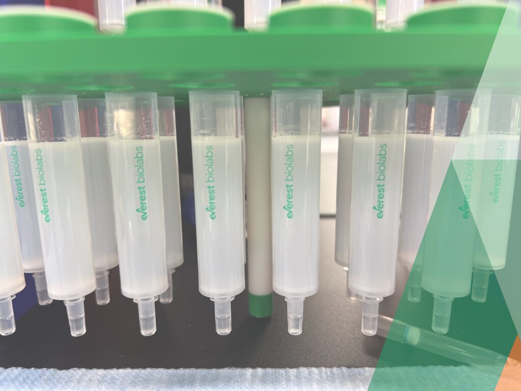
Why is SEC becoming the most widely used EV isolation technique?
Why is SEC becoming the most widely used EV isolation technique?
Read MoreReferences
High-Throughput Extracellular Vesicle Isolation Using Plate-Based Size Exclusion Chromatography and Automation pubs.acs.org/doi/10.1021/jacs.4c17948
This study introduces a plate-based SEC workflow for reproducible, automated, high-throughput extracellular vesicle isolation from plasma and other biofluids.
Read more
The authors adapted size exclusion chromatography (SEC) into a 24-well plate format, where each resin-packed well functions as an individual SEC column. Using EV markers CD63 and CD81, along with albumin as a marker of free-protein contamination, they optimized separation conditions and demonstrated that the workflow can be automated using liquid-handling platforms. This approach addresses a major bottleneck in EV biomarker research by enabling scalable, reproducible EV isolation across large sample sets, with the potential to process hundreds of samples per day for biomarker discovery and diagnostic applications.
Framework for rapid comparison of extracellular vesicle isolation methods. eLife 2021 doi.org/10.7554/eLife.70725
This study presents a standardized framework for assessing the efficiency and purity of different extracellular vesicle (EV) isolation techniques.
Read more
The authors utilized ultrasensitive single-molecule array (Simoa) assays to quantify three key EV transmembrane proteins-CD9, CD63, and CD81- while measuring albumin levels as a marker of free protein contamination. By applying this approach to plasma and cerebrospinal fluid (CSF), they systematically compared commonly used isolation methods, including ultracentrifugation, precipitation, and size exclusion chromatography (SEC). The results highlight SEC as a superior method for maintaining both yield and purity, particularly when optimized with custom column parameters. This study provides a valuable, reproducible strategy for improving EV isolation, aiding biomarker discovery and translational research in EV-based diagnostics.
Improved isolation of extracellular vesicles by removal of both free proteins and lipoproteins. eLife 2023 doi.org/10.7554/eLife.86394
This study presents an advanced method for isolating extracellular vesicles (EVs) from plasma by minimizing contamination from free proteins and lipoproteins, which traditionally complicate EV purification.
Read more
The researchers developed a digital ELISA assay targeting ApoB-100, a key lipoprotein marker, and integrated it with existing assays for albumin and EV-associated tetraspanins. They systematically evaluated various size exclusion chromatography (SEC) resins and developed a novel approach, Tri-Mode Chromatography (TMC), to enhance EV purity while maintaining yield. The study highlights the advantages of TMC in reducing co-isolated contaminants and improving the reliability of EV-based biomarker discovery, particularly for proteomics applications.
Measurement of α-synuclein as protein cargo in plasma extracellular vesicles. PNAS 2024 doi.org/10.1073/pnas.2408949121
This study addresses the challenge of measuring α-synuclein, a key protein associated with Parkinson’s disease (PD), within extracellular vesicles (EVs) isolated from plasma.
Read more
Given the difficulty of distinguishing EV-associated proteins from free plasma proteins, the researchers developed a method combining optimized size-exclusion chromatography (SEC) for EV isolation with a protease protection assay and ultrasensitive digital ELISA (Simoa) measurements. Their analysis revealed that only a small fraction of total plasma α-synuclein is contained within EVs, but its phosphorylated form (pSer129), a marker of PD pathology, is enriched within EVs compared to free plasma protein. Applying this method to patient samples, they observed subtle but significant differences in EV α-synuclein and pSer129 levels between PD, Lewy body dementia (LBD), and control groups. This work establishes a robust framework for studying EV-contained neurodegenerative biomarkers and highlights the potential of EV-based diagnostics for neurodegenerative diseases.
Identification of markers for the isolation of neuron-specific extracellular vesicles. bioRxiv 2024 doi.org/10.1101/2024.04.03.587267
This study presents a systematic approach for identifying neuron-derived extracellular vesicle (EV) markers, facilitating the selective isolation of neuron-specific EVs from cerebrospinal fluid (CSF) and plasma.
Read more
Researchers developed a framework that integrates gene expression data with EV proteomics to identify transmembrane proteins unique to neurons. They optimized high-purity EV isolation by combining multiple purification techniques, including size exclusion chromatography (SEC), density gradient centrifugation (DGC), and Mixed Mode Resin (MMR) Slurry, to effectively remove free proteins and lipoprotein contaminants while preserving EV integrity. Through proteomic analysis, they identified NRXN3 as a robust neuron-specific EV marker and validated its presence using ultrasensitive immunoassays. By optimizing immuno-isolation protocols, the study provides a foundation for isolating neuron-derived EVs, enabling their use in biomarker discovery for neurological diseases. This methodology offers a scalable strategy for isolating cell type-specific EVs, expanding potential applications in liquid biopsy and neurodegenerative disease diagnostics.
